
Spinal Deformity & Scoliosis
Three-dimensional correction guided by global balance and patient-specific alignment, with deliberate decisions about what movement can be preserved.
Clinical expertise
My practice includes lumbar and cervical conditions, scoliosis and complex reconstruction. Experience with different approaches allows treatment to follow the diagnosis, the anatomy and what you want it to achieve.
Three-dimensional correction guided by global balance and patient-specific alignment, with deliberate decisions about what movement can be preserved.
Reassessing persistent symptoms, instability or complications after previous operations, and planning revision surgery when further intervention is justified.
Treating conditions of the lower back and neck, including nerve compression and instability, with decompression, fusion or reconstruction when needed.
Understanding how the spine and pelvis work together in standing and movement, particularly before deformity correction or a larger reconstruction.
Considering cervical disc replacement where movement can be preserved safely, and endoscopic techniques where they provide suitable access to the problem.
Using an approach from the front, including ALIF, when the anatomy and reconstruction goals make it appropriate.
Selected surgical cases
Scoliosis · A 17-year-old patient
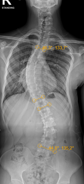
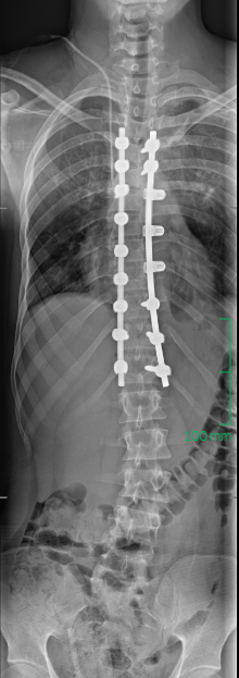
The lower back remains unfused.
Select an image to enlarge it in a new tab. Images are shown at different magnifications.
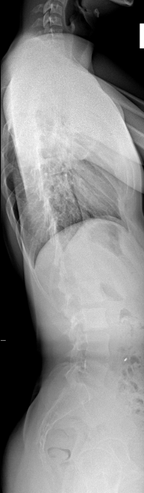
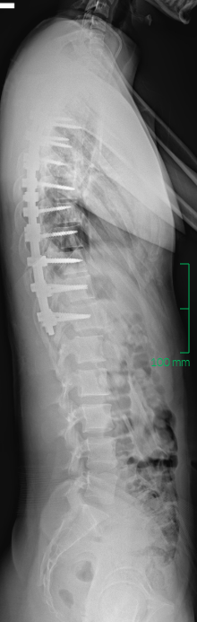
A scoliosis operation is not chosen from the Cobb angle alone. I ask how much growth remains, which curves are structural or flexible, how the trunk and pelvis are balanced, whether a leg-length difference is influencing the standing X-ray, and what movement the patient wants to keep.
Only then do I decide where the fixation should start and stop. I examine each vertebra closely; if a pedicle is too small or a screw path is unsafe, another fixation method can be used. In this case, sublaminar bands provided that alternative, allowing a selective thoracic fusion from T4 to T12.
The aim is not simply a straighter X-ray. It is a balanced correction that leaves the lumbar spine free to move while considering the effect of the leg-length discrepancy when assessing the remaining lumbar curve.
When I plan surgery, the correction on the X-ray and the function that remains both matter.
Adult scoliosis · Degenerative change
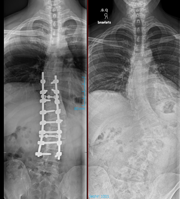
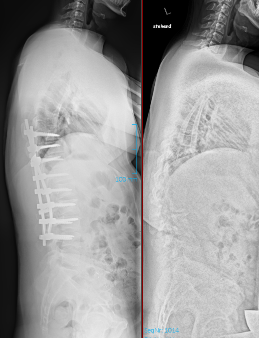
A longstanding scoliosis had become stiff and rotated, with degenerative changes superimposed. A straighter frontal X-ray alone would not define a good plan.
I considered the three-dimensional curve, the affected levels and the standing profile from both front and side. These factors shaped the correction and where the fusion should begin and end.
Here the priorities were improved alignment and durable stabilisation. The postoperative radiographs show the correction and fixation. Follow-up assesses pain, movement and the ability to manage daily activities.
Different curves call for different goals. The plan must fit the patient's anatomy and the function we are trying to protect.